
Researchers at Osaka University have demonstrated, for the first time, that lysosomes, when damaged, undergo a repair process known as microautophagy. This mechanism plays a crucial role in hindering the aging process.
Aging is a fundamental process that affects organisms at the cellular level. As cells age, they encounter both self-inflicted damage and external harm. To combat this, cells rely on various mechanisms to maintain their health and stability.
One key player in this process is the lysosome, a critical cellular structure responsible for digesting damaged components and pathogens. This helps maintain cellular and tissue stability. However, an intriguing question arises: can lysosomes themselves be repaired when damaged, and if so, what are the mechanisms behind this repair?
Microautophagy: The Key to Lysosomal Repair
In a study recently published in EMBO Reports, researchers from Osaka University and Nara Medical University have shown that damaged lysosomes are repaired by a mechanism called “microautophagy” and have identified two key regulators of this process.
Microautophagy is one of the three main types of autophagy in most higher organisms. It is a regulated process by which cellular components that have become dysfunctional or are no longer required are broken down. Although it is assumed to be involved in defense mechanisms collectively called lysosomal damage responses, the details remain unknown.
Lysosomes frequently become damaged and lysosomal dysfunction has been linked to accelerated aging and a shortened lifespan. In this study, the researchers tried to understand the repair mechanisms. To identify a novel regulator of lysosomal damage response, they focused a signaling pathway called Hippo pathway which controls multiple processes such as cellular growth.
They knocked down individual components of the Hippo pathway in the human cells, and then observed whether the cells could respond to induced lysosomal damage. This screening revealed that a protein called Serine-threonine kinase 38 (STK38) is essential for the lysosomal damage response.
Discovering the Mechanisms of Lysosomal Repair
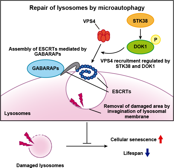
They then found that STK38 works with a protein complex called the “endosomal sorting complex required for transport (ESCRT) machinery”, which was already known to be linked to lysosomal repair.
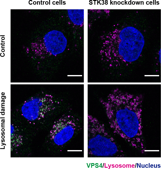
“STK38 recruits the protein ‘vacuolar protein sorting 4’ (VPS4) to damaged lysosomes and is crucial for disassembling the ESCRT machinery at the end of the repair process,” explains lead author of the study Monami Ogura. They further found that lysosomal membrane repair by ESCRT machinery is mediated by microautophagy.
The Role of GABARAPs in Lysosomal Repair
They also identified that non-canonical lipidation of a subfamily of autophagy-related protein 8 (ATG8s) molecules—the key autophagy proteins—known as Gamma-aminobutyric acid receptor-associated proteins (GABARAPs) is required for this process. Lipidation, the process of modifying ATG8s with lipid extensions, is the main process involved in autophagy. In non-canonical lipidation ATG8s are lipidated into single-membrane endolysosomes, instead of double-membrane phagophore seen in canonical lipidation.
The researchers showed that the GABARAPs are essential for the first step of the process of lysosomal repair. “We showed that non-canonical lipidation of ATG8s is crucial for the initial recruitment of the ESCRT machinery to damaged lysosomes and their subsequent repair,” explains senior author Shuhei Nakamura.
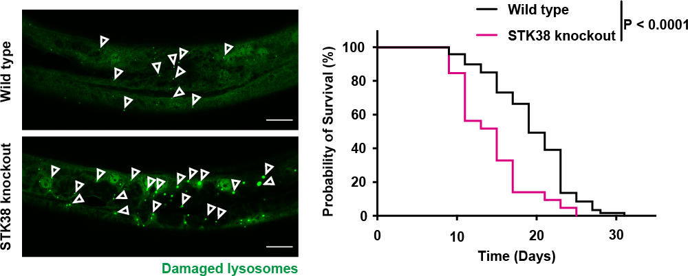
The team showed that depletion of the regulators of microautophagy increased the rate of senescent cells and shortened lifespan in C. elegans. Both STK38 and GABARAPs also have evolutionarily conserved roles, indicating the significance of this pathway in maintaining lysosomal integrity, healthy cellular function, and the prevention of cellular senescence and organismal aging. The detailed understanding provided by this study paves the way for increasing healthy aging and has great therapeutic value for the treatment of age-related diseases.
The study was funded by the Japan Society for the Promotion of Science, the Ministry of Education, Culture, Sports, Science and Technology, the Japan Agency for Medical Research and Development, and the Japan Science and Technology Agency.